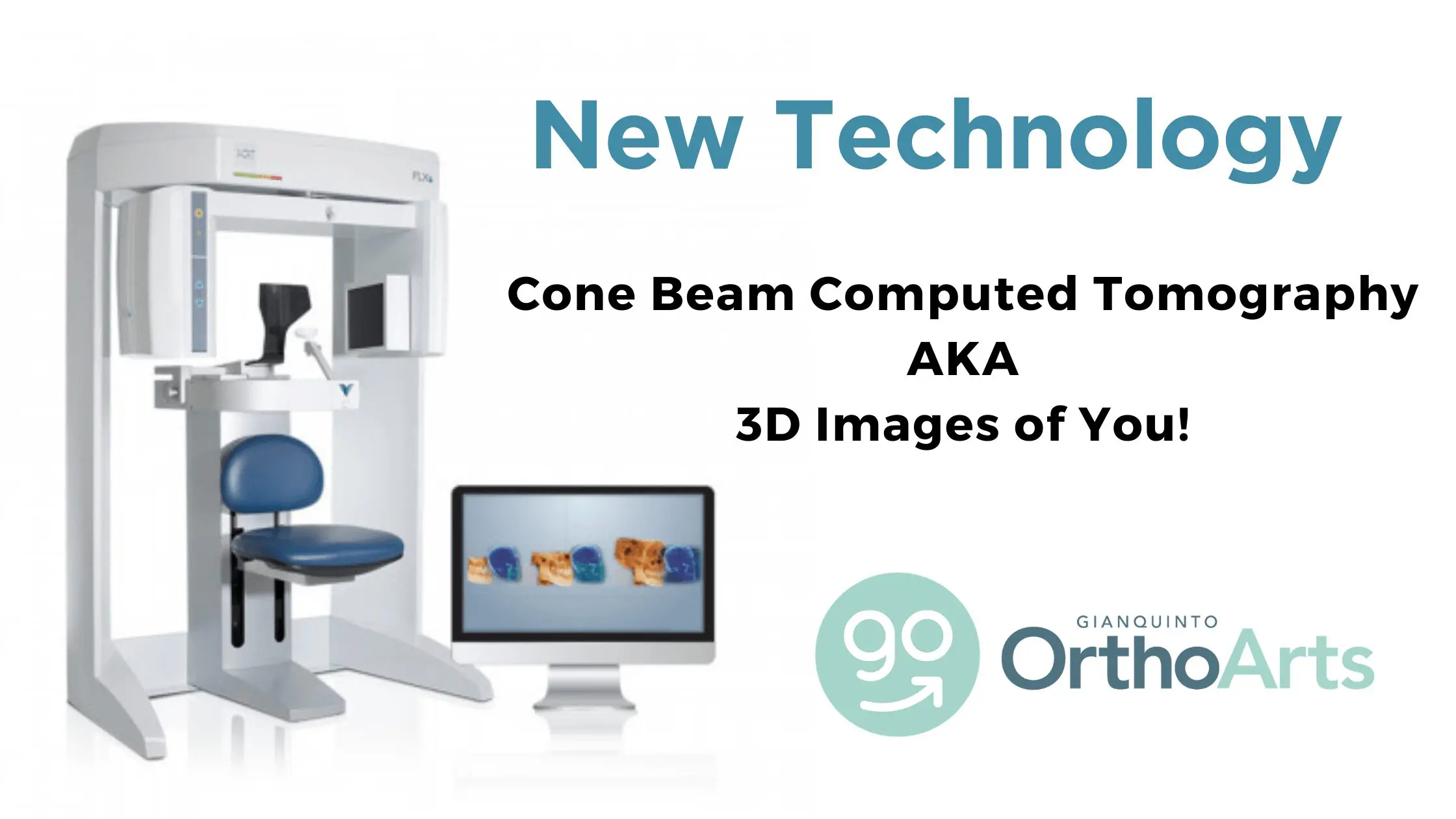
What is Cone Beam 3D Imaging?
Traditionally, orthodontic diagnosis and treatment planning has been done with 2-dimensional imaging using a panoramic x-ray, generally showing what teeth are present and where they are, and a cephalometric x-ray, which is a side view that gives a general idea of jaw positioning. Cone Beam Computed Tomography, or CBCT, is a 3D x-ray that shows so much more information in a single image, with much higher accuracy than ever before.
Why did Dr. G invest in Cone Beam Computed Tomography for his patients?
2D imaging is still considered the base level standard of care, but we’ve always wanted to provide the best. When our 2D x-ray machine needed replacement, we upgraded to the best-in-class, more technologically advanced 3D imaging.
How does Cone Beam 3D Imaging Work?
Instead of taking several 2D images, each requiring exposure to radiation to a fan-shaped beam, the x-ray generator produces a cone-shaped beam that allows the sensor to capture 3D information to recreate any 2D or 3D image we need.
What difference do 3D images make?
Remember the days of paper maps? If you had access to google maps with satellite photos and live traffic, would you still use a paper map? Or look at the sun to tell time? Probably not. It’s the same with 3D x-rays vs. 2D. We can do a much better job of treatment planning and no longer have to make educated anatomical guesses. When we plan our cases for treatment with custom digital appliances, 3D imaging is an absolute must. It’s the difference between knowing and guessing. That means faster, safer, and much more comfortable treatment.
What are the benefits of Cone Beam 3D imaging?
Dental cone beam computed tomography (CT) is a special type of x-ray equipment used when regular dental or facial x-rays are insufficient. Your doctor may use this technology to produce three-dimensional (3-D) images of your teeth, soft tissues, nerve pathways, and bone in a single scan.
- The focused x-ray beam reduces scatter radiation, resulting in better image quality.
- A single scan produces a wide variety of views and angles that provide a complete evaluation.
- Cone beam CT scans provide more information than conventional dental x-ray, allowing for more precise treatment planning.
- CT scanning is painless, non-invasive, and accurate.
- A major advantage of CT is its ability to image bone and soft tissue at the same time.
- No radiation remains in a patient’s body after a CT examination.
- X-rays used in CT scans should have no immediate side effects.
These images are our standard of care and taken whenever needed before or during orthodontic treatment. The patient is only charged if a physical copy is needed for a referral to another specialist for treatment such as surgery, implants, airway evaluation, or other purposes. Our referring dentists have access to the x-rays of all of their patients under our care.
Specific case examples:
Let’s compare the 2D and 3D images in the cases below. You will be able to see the benefits of 3D imaging.
Case 1: Need for extractions. In the 2D x-rays, it looks like there’s plenty of room to fit all of the teeth in the bone. But when looking at the case in 3D, it becomes obvious that the roots of several teeth are already outside the bone, and treating the case without removing teeth would only make this worse.


Case 2: Pathology. This patient came in for a routine consultation, and we noticed something that didn’t look right. On the 2D x-ray, the dark spot doesn’t look too significant. But when looking at the case in 3D, you can see a large area of bone is missing due to a long-standing infection that his general dentist hadn’t diagnosed. We were able to refer the patient to another specialist for evaluation and treatment.


Case 3: Extra teeth. His general dentist referred this patient because his right front tooth wasn’t coming in. The 2D x-ray doesn’t look too out of the ordinary, but in the 3D view, we can see a supernumerary (extra) tooth blocking the eruption of the one we are expecting to see. We were able to refer this patient to an oral surgeon to remove the extra tooth and normalize the eruption pattern.


This is just another example of how our hometown veteran orthodontist Jared Gianquinto goes the extra mile to provide the gold standard of treatment for his patients right here in Bakersfield. Are you interested in learning more about what your options are for a smile of a lifetime? Be sure to call our office at 661-829-7970 or book a virtual consultation right here, you have nothing to lose since it’s totally complimentary!